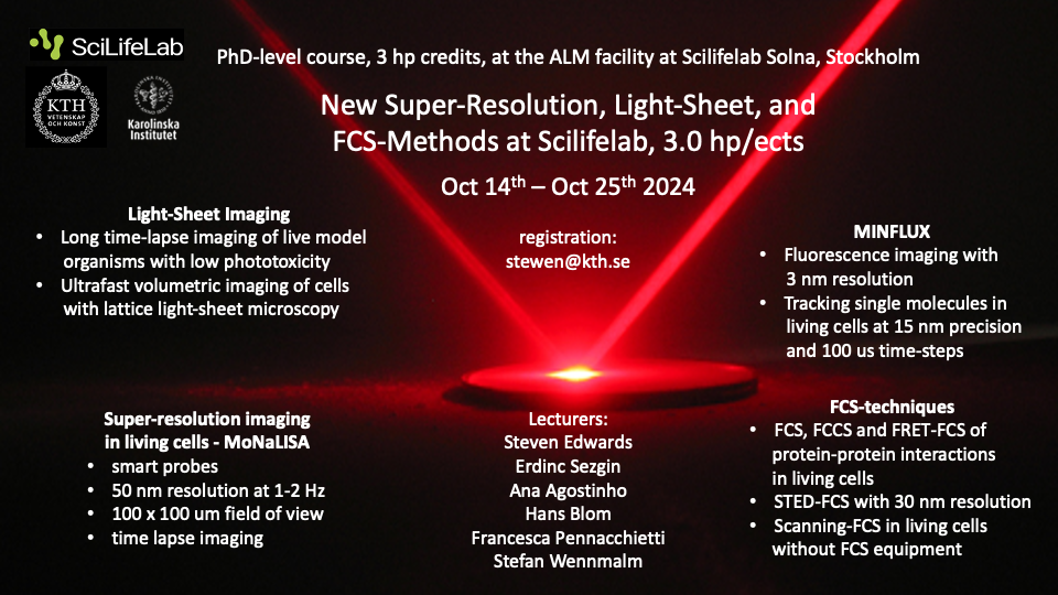
The Advanced Light Microscopy (ALM) SciLifeLab node of NMI gives a two-week PhD-level course on four new imaging and fluorescence spectroscopy techniques: Depletion-based Super-Resolution Imaging, MINFLUX, Light-Sheet Imaging, and FCS-Methods. Do not miss this opportunity if you are interested in learning about bioimaging, from molecules up to live model organisms, using advanced light microscopy.
Dates: Monday October 14 – Friday October 25, 2024 Credits: 3 hp for PhD students Location: Gamma 3, SciLifeLab, Solna Registration deadline: September 30, 2024 Registration: stewen@kth.se
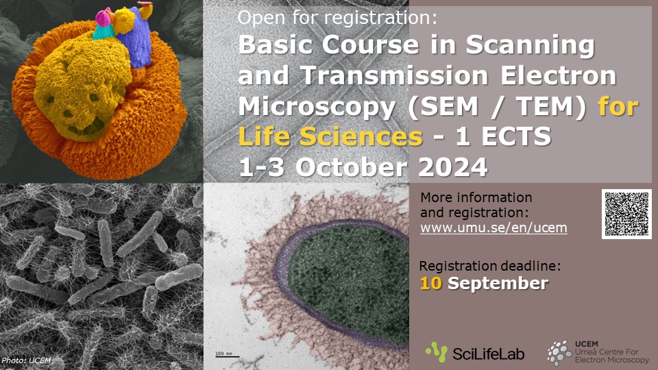
Welcome to this electron microscopy course at our NMI node UCEM in Umeå. The course covers lectures and practical demonstrations in SEM and TEM techniques. The contents include principles of electron microscopy, specimen preparation, cryo-electron microscopy and correlative light-electron microscopy. The main focus will be on imaging biological samples, but the course may also be suitable for material scientists interested in high-resolution electron microscopy.
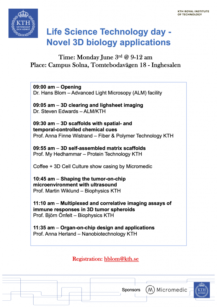
The Advanced Light Microscopy (ALM) NMI node at SciLifeLab welcomes you to a KTH networking event where leading KTH scientists present novel 3D biological sample generation and visualisation applications.
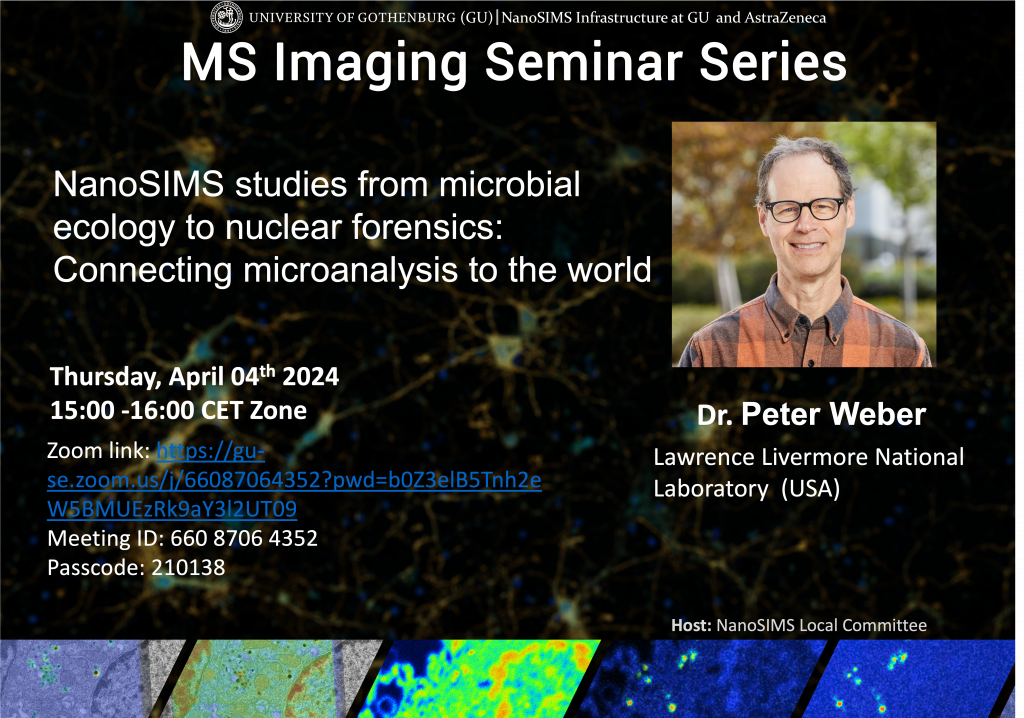
Take the chance to join this Mass Spectrometry Imaging Zoom seminar on the 4th April, 15:00-16:00 (CET time). It will be given by Dr. Peter Weber from Lawrence Livermore National Laboratory, US. Dr. Weber is an expert in NanoSIMS imaging, as well as multiphoton microscopy and coherent Raman imaging. His research focus is microanalysis, microbial ecology, soil processes, elemental and isotopic tracers, and microbial and nuclear forensics. He has analyzed a broad range of materials, worked on methods to link microbial function and identity in complex microbial communities, and has studied microbial mineralization, symbiosis, predation, nitrogen fixation, carbon fixation, carbon cycling and phosphorus uptake. Dr. Weber is interested in correlated imaging and pushing the limits of compositional sensitivity, including developing methods to analyze viruses, cell membranes, and microarrays.
Title: “NanoSIMS studies from microbial ecology to nuclear forensics: Connecting microanalysis to the world”
The ZOOM seminar will be held 15:00-16:00 including discussion, then 16:00-16:30 will be a separate informal discussion time for those that want to stay.
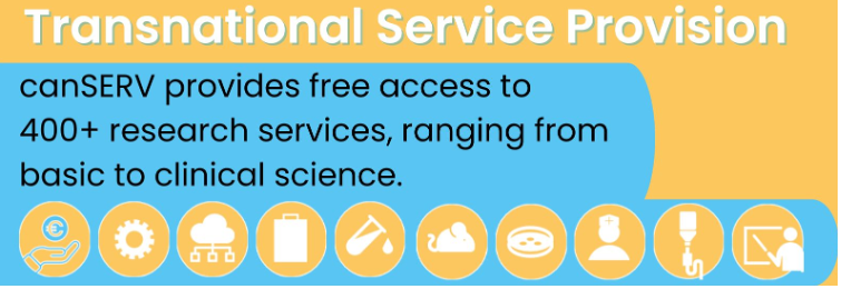
Euro-BoImaging is pleased to announce that the second Open Call from the Horizon Europe-funded canSERV project is here! All user projects – ranging from basic discovery science to translational science and into personalised oncology on any type of cancer – are eligible. The total indicative funding volume of this call is 1 Million Euro across the entire canSERV consortium. Within this project, NMI and many other Euro-Bioimaging Nodes offer access to their expertise. Read more in the links below!
Welcome to a practical course on sample preparation for TEM at the NMI node UCEM in Umeå. Participants will practice TEM sample preparation and TEM imaging with a sample from their own project. The course includes Ultra-microtome sectioning, Chemical fixation, Plastic embedding, TEM imaging, High-pressure freezing & freeze substitution, Immunolabeling techniques (Immunogold, CLEM) and Volume EM (FIB-SEM & Tomography). For more information, please visit the course website, see link below.
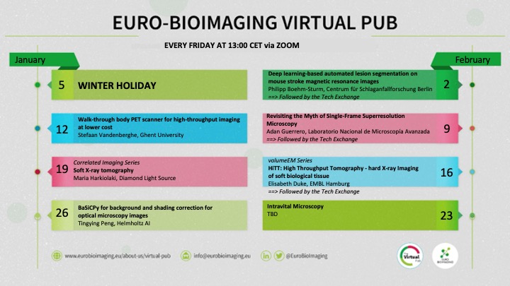
Did you know that Euro-BioImaging has a Virtual Pub every Friday at 13:00 CET on Zoom? Node staff and friends of Euro-BioImaging join with the Euro-BioImaging Hub staff for a virtual meeting – about topics that interest us all. We showcase our Nodes’ success stories and expertise, exciting science, travel grants, new technologies – and more!
Well, now the Virtual Pub is on Winter Break until January 12, 2024 but please register for the 2024 season by following the link below. Above you can see the schedule for January and February. Everyone is welcome!
Euro-BioImaging is pleased to announce that the first Open Call from the Horizon Europe-funded canSERV project is here! Cancer Researchers are invited to apply for FREE state-of-the-art services and training at several European Research Infrastructures, including Euro-BioImaging ERIC. Within this project, NMI and many other Euro-Bioimaging Nodes offer access to their expertise. It’s an amazing opportunity for the cancer research community to access a wide-ranging portfolio of services.
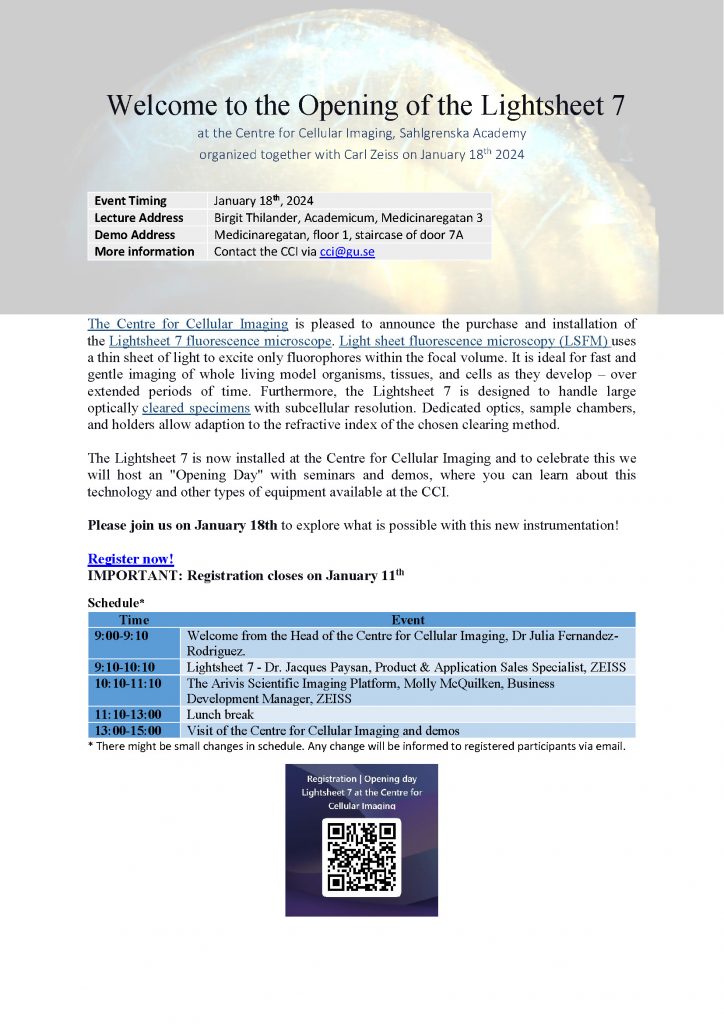
The Centre for Cellular Imaging (CCI), our NMI node in Gothenburg, has installed a new Lightsheet 7 fluorescence microscope and will celebrate this by hosting an opening day with seminars and demos of both the new Lightsheet instrument and other equipment at CCI. Please join us on January 18th to explore what is possible with this new instrumentation!
Organizer: CCI Sahlgrenska Academy & Zeiss Location for lecture: Birgit Thilander, Academicum, Medicinaregatan 3, Göteborg Location for demo: Medicinaregatan, floor 1, staircase of door 7A, Göteborg Time of event: January 18, 2024 Deadline for registration: January 11, 2024 Registration weblink: https://forms.office.com/e/YH4eYMYSSy Contact: cci@gu.se
See the flyer below for more information!
This website uses cookies! By continuing to use this site, you accept our use of cookies. They help feed our microscopes!