This year the upcoming SMLMS 2024 symposium, a prestigious gathering of leading scientists and researchers in the field of single-molecule localization microscopy and advanced super-resolution imaging technologies, celebrates the 10th anniversary of groundbreaking advancements since the Nobel Prize was awarded for developing super-resolved fluorescence microscopy. Keynote speaker is Nobel Prize laureate in Chemistry Professor W. E. Moerner from Stanford University, USA. For more info and registration, visit the website.
Time: August 28th to 30th, 2024 Location: Lisbon, Portugal Website: https://2024.smlms.org
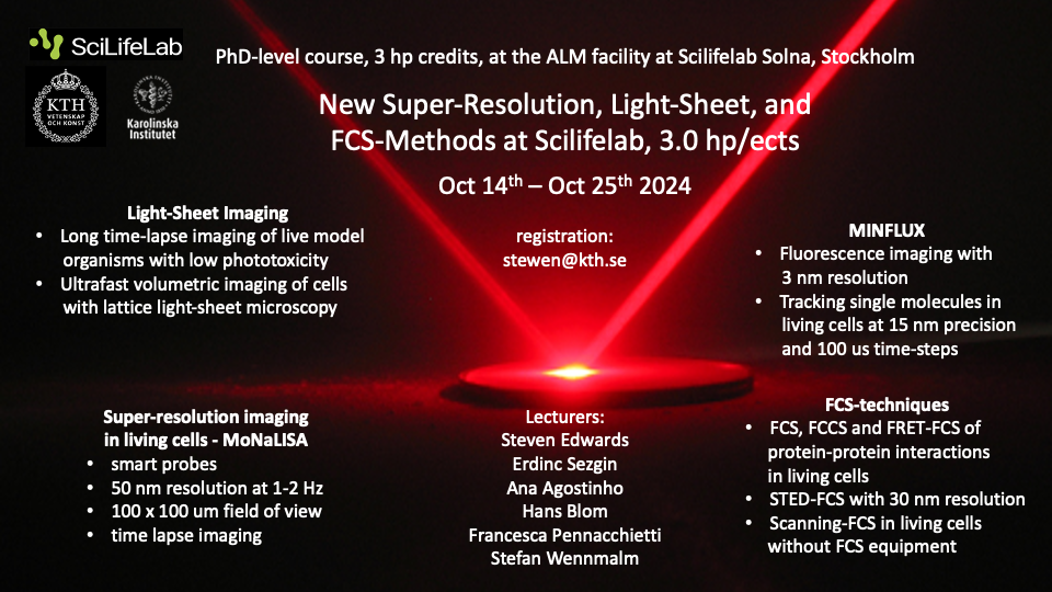
The Advanced Light Microscopy (ALM) SciLifeLab node of NMI gives a two-week PhD-level course on four new imaging and fluorescence spectroscopy techniques: Depletion-based Super-Resolution Imaging, MINFLUX, Light-Sheet Imaging, and FCS-Methods. Do not miss this opportunity if you are interested in learning about bioimaging, from molecules up to live model organisms, using advanced light microscopy.
Dates: Monday October 14 – Friday October 25, 2024 Credits: 3 hp for PhD students Location: Gamma 3, SciLifeLab, Solna Registration deadline: September 30, 2024 Registration: stewen@kth.se
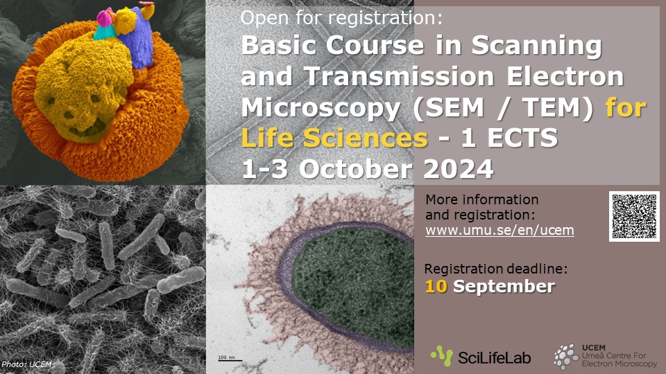
Welcome to this electron microscopy course at our NMI node UCEM in Umeå. The course covers lectures and practical demonstrations in SEM and TEM techniques. The contents include principles of electron microscopy, specimen preparation, cryo-electron microscopy and correlative light-electron microscopy. The main focus will be on imaging biological samples, but the course may also be suitable for material scientists interested in high-resolution electron microscopy.
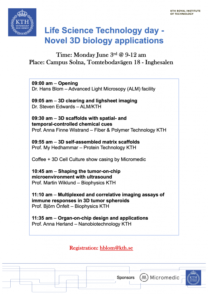
The Advanced Light Microscopy (ALM) NMI node at SciLifeLab welcomes you to a KTH networking event where leading KTH scientists present novel 3D biological sample generation and visualisation applications.
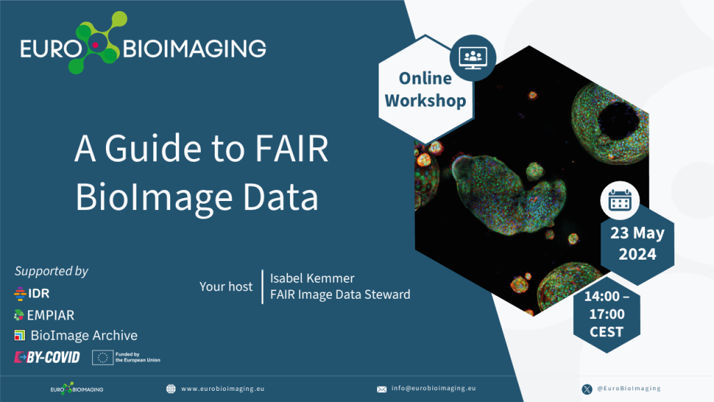
If you perform biological imaging and want to maximise the potential of your bioimaging data, we suggest you to take a look at this free workshop, Euro-BioImaging’s Guide to FAIR Bioimage Data, provided by Euro-BioImaging. In this interactive online workshop you will learn about the FAIR principles and benefits in the context of bioimaging data, best practices for data management and much more. There is also an opportunity to bring your own data to the workshop. Read more by clicking the links below.
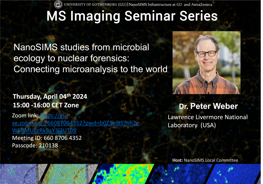
Take the chance to join this Mass Spectrometry Imaging Zoom seminar on the 4th April, 15:00-16:00 (CET time). It will be given by Dr. Peter Weber from Lawrence Livermore National Laboratory, US. Dr. Weber is an expert in NanoSIMS imaging, as well as multiphoton microscopy and coherent Raman imaging. His research focus is microanalysis, microbial ecology, soil processes, elemental and isotopic tracers, and microbial and nuclear forensics. He has analyzed a broad range of materials, worked on methods to link microbial function and identity in complex microbial communities, and has studied microbial mineralization, symbiosis, predation, nitrogen fixation, carbon fixation, carbon cycling and phosphorus uptake. Dr. Weber is interested in correlated imaging and pushing the limits of compositional sensitivity, including developing methods to analyze viruses, cell membranes, and microarrays.
Title: “NanoSIMS studies from microbial ecology to nuclear forensics: Connecting microanalysis to the world”
The ZOOM seminar will be held 15:00-16:00 including discussion, then 16:00-16:30 will be a separate informal discussion time for those that want to stay.
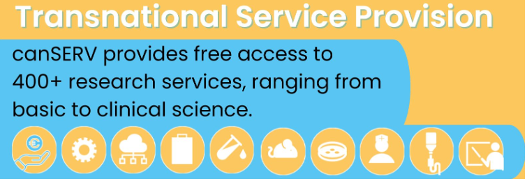
Euro-BoImaging is pleased to announce that the second Open Call from the Horizon Europe-funded canSERV project is here! All user projects – ranging from basic discovery science to translational science and into personalised oncology on any type of cancer – are eligible. The total indicative funding volume of this call is 1 Million Euro across the entire canSERV consortium. Within this project, NMI and many other Euro-Bioimaging Nodes offer access to their expertise. Read more in the links below!
We look forward to the next Euro-BioImaging User Forum – this time on the topic of IMAGE DATA. The User Forum will take place on March 26th, 2-5 pm CET. This is a great opportunity to highlight the innovative image analysis and image data management solutions developed by you all at the Euro-BioImaging Nodes and supporting Euro-BioImaging Users!
A selection will be made among the submitted abstracts for joint presentations from Users and Nodes that should cover both scientific aspects of the research project as well as the technical aspects of the work. While presentations from Euro-BioImaging users are encouraged, other users of Euro-BioImaging Node facilities are also invited to submit their abstracts. So please consider if you have an interesting story from your users to share.
Welcome to a practical course on sample preparation for TEM at the NMI node UCEM in Umeå. Participants will practice TEM sample preparation and TEM imaging with a sample from their own project. The course includes Ultra-microtome sectioning, Chemical fixation, Plastic embedding, TEM imaging, High-pressure freezing & freeze substitution, Immunolabeling techniques (Immunogold, CLEM) and Volume EM (FIB-SEM & Tomography). For more information, please visit the course website, see link below.