We welcome you to two symposiums in Stockholm on optical clearing and expansion microscopy. The first symposium takes place at the Wenner-Gren Center on Sep 12th, and the second symposium at SciLifeLab on Sep 13th-14th. Please see the below flyers for more info.
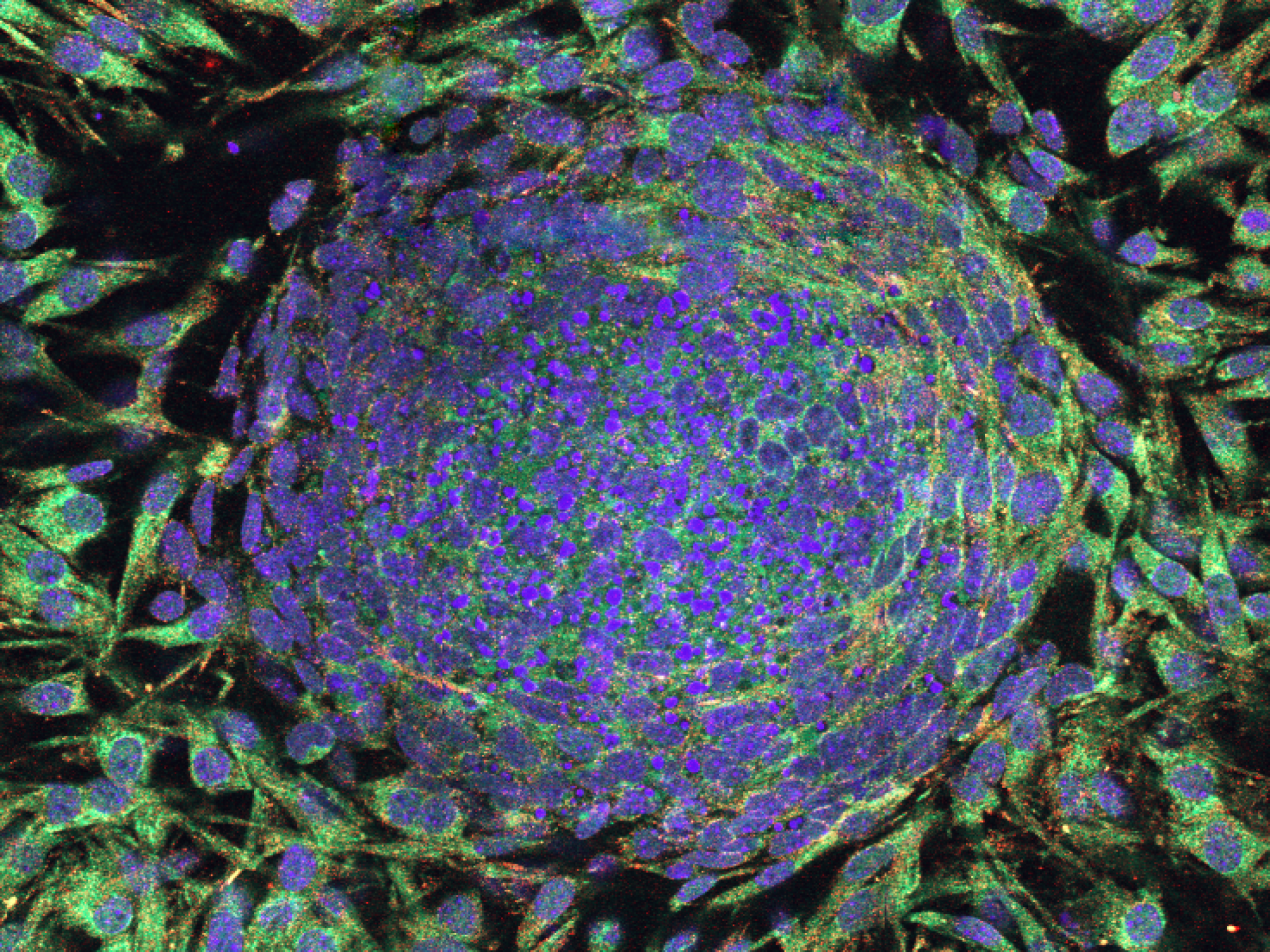
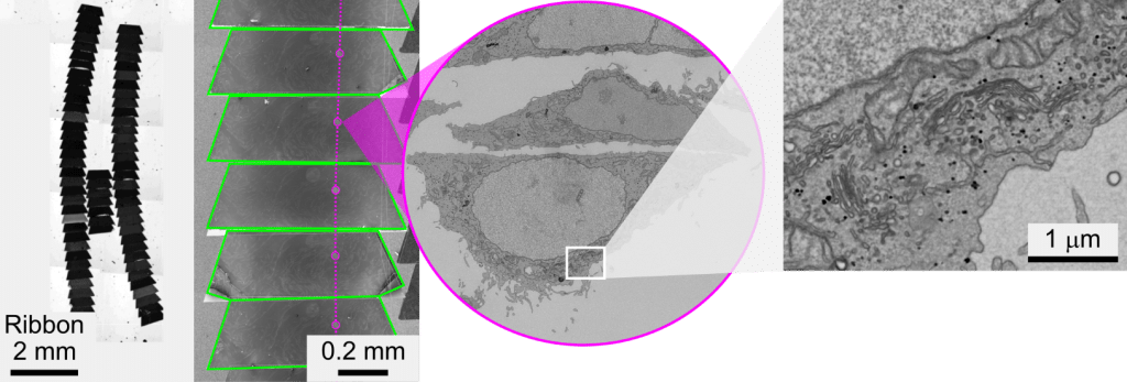
The Centre for Cellular Imaging, which is the national SciLifeLab facility for Correlative Array Tomography in Sweden, has the great pleasure to welcome you to the first Cellular 3D volume – Correlative Array Tomography (CAT) Workshop! It is designed to bring experts, researchers and industry together interested in Cellular 3D volume – CAT to introduce this new technology, stimulate knowledge exchange and the formation of collaborations.
This advanced course on methods in bioimage analysis concentrates on teaching cutting-edge concepts and tools for quantitative image analysis and will seek to upgrade the competencies of future bioimage analysis experts on both theoretical algorithm advancements as well as on practical implementation skills.
The registration deadline for the DIASA course has been prolonged to March 25th, click here for more info. This is great opportunity to learn more about image analysis for scientific applications. Go to the course homepage and sign up!
This course covers lectures and practical demonstrations in super-resolution microscopy and light-sheet imaging, as well as fluctuation correlation spectroscopy techniques. During week 2 participants (at home) use the aquired knowledge about the advance microscopy techniques to plan their own research projects.
Organizer: KTH-ALM Location: SciLifeLab in Solna, Tomtebodavägen 23 Time: May 22nd-25th lectures/demos; May 29th – June 2nd own project, 2023 Contact and registration: stewen@kth.se, Stefan Wennmalm KTH/SciLifeLab
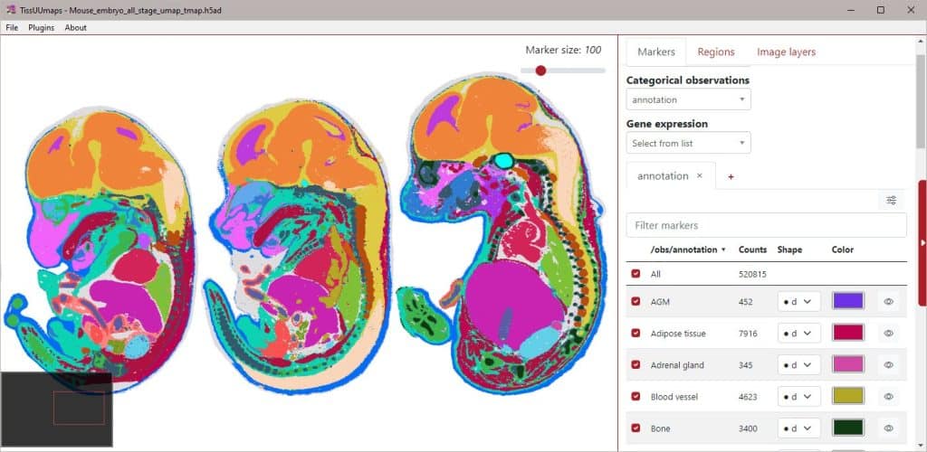
We present TissUUmaps, browser-based tool for GPU-accelerated visualization and interactive exploration of tens of millions of datapoints overlaying tissue samples. Users can visualize markers and regions, explore spatial statistics and quantitative analyses of tissue morphology, and assess the quality of decoding in situ transcriptomics data. TissUUmaps provides instant multi-resolution image viewing, can be customized, shared, and also integrated in Jupyter Notebooks. It is also possible to directly connect spatial markers with markers in feature space, such as UMAP plots, to interactively relate feature space with physical space. TissUUmaps was created in collaboration between BIIF and the Wählby lab. You can read more about it and test the software on its web page: https://tissuumaps.github.io/
During the seminar, we will specifically showcase new features of TissUUmaps 3.1, such as: – HDF5 / AnnData files loading – Network diagram visualization – Multiple datasets displayed on a grid – Plugin engine
The webinar will be given by Christophe Avenel and Fredrik Nysjö.
The BioImage Informatics Facility (BIIF) together with microscopy expert Sylvie Le Guyader (LCI, Karolinska Institutet) organizes a Call4Help session every month. The aim is to offer combined expertise towards microscopy and bioimage analysis. All researchers from Swedish institutes can participate.
NMI node: BIIF at Uppsala University Location: on zoom Time of event: every 1st week of the month (mainly on Tuesdays, but there might be exceptions) Deadline for registration: continuous Weblink: https://www.scilifelab.se/call4help-form/ Contact email: biif@it.uu.se